Blog

Thu Dec 05 2019
Retinal Detachment – A Flash is all that it takes!
Read More
Retinal Detachment – A Flash is all that it takes!
Thu Dec 05 2019
Retinal Detachment – A Flash is all that it takes!

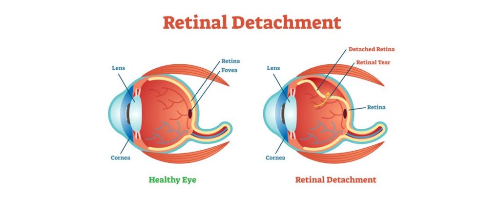
Imagine it’s just a regular day and you are doing your daily chores, suddenly, you see a flash of light and maybe some floaters. You may feel a little nauseous instantly and then there are chances that you do nothing about it. Much later, you may feel a blurry vision. One may panic at this point. A good eye consultation is required without any delay. Such symptoms could be of retinal detachment. Read this piece of information to understand more. It is a detachment or tearing of the retina from its supporting tissues that provide the retina with oxygen nourishment and further connects the received image to the brain. It implies an emergency where one has to immediately rush to the hospital as the chances of partial or total vision loss is expected. Sudden flashes of light, spots, or multiple floaters are the most basic symptoms of retinal detachment. One may start having blurry visions or feel a curtain-like shadow over the visual field; even the peripheral vision starts diminishing. Please note that the aforementioned primary symptoms are painless. You may not experience any pain and may get away from the situation, thinking it’s nothing important, but if left unattended, partial or complete vision loss is the final destination. It’s also important to note the type of retinal detachment that one may experience; there are three types-

  • Rhegmatogenous Retinal Detachment-

Being the most common type of retinal detachment, it occurs through a hole in the retina. It results in the flow of fluid from within the eye to get behind the back of the retina. This interrupts the supply of oxygen and essential nutrients that are received by the retina.

  • Tractional Retinal Detachment-

Familiar among people with a poorly managed diabetes-this type of detachment occurs when the scar tissue of the retina contracts and pulls away from the cells and tissues that are connected with the retina.

  • Exudative Retinal Detachment-

Abnormal development, lack of protein, cancer behind the retina, inflammation, or any fluid accumulation in the back of the eye results in Exudative Retinal Detachment.

Other reasons for retinal detachment are nearsightedness and at times even after LASIK surgery, cataract surgery, tumors, or other eye diseases may cause the retina to pull away. The risk of retinal detachment also increases among people who have a history of retinal detachment running in their family, or they are over 50 years of age, etc. But since retinal detachment can occur anytime and in anyone, with or without any prior warning or history, what happens once you have rushed to the hospital and waiting for treatment to bring back your complete vision? The sooner the retina is attached back, the better it is. Retinal specialists are the ones who engage in this type of surgery. Depending on the type and cause, the following retinal detachment treatments are provided-

  • Scleral Buckling Surgery –

Being the most common type of surgery in case of retinal detachment, the procedure consists of putting up a small piece of a band made of plastic or silicon on the sclera of the eye. The band helps in compressing the eye inwardly and allowing the retina to attach internally.

  • Vitrectomy –

In this type of surgery, the posterior chamber of the eye that is the clear jelly-like substance is removed from the eye and is replaced with a clear silicon oil that helps in pushing back the detached retina into its place.

  • Pneumatic Retinopexy –

In this method, a small bubble of gas is introduced in the vitreous body of the eye that works to push the detached retina back in its space.

Other treatments also include fixing the retina back through the laser method, which is also called cryopexy. However, surgical reattachments depend a lot on factors like location, cause, the extent of detachment, etc. that impact the success of the surgery. The recovery post-treatment may take 3 to 6 weeks. Depending on the nature of your treatment, the doctor would provide you with a list of instructions to be followed during the recovery period. However, there is no known way of preventing retinal detachment from occurring, but if you are struggling with the thought of being prone to retinal detachment, then here are some quick ideas that you can adapt to skip the risk of the condition that is retinal detachment-

  • Wear protective glasses when playing sports or other activities to avoid any injury
  • Make a constant effort to control the diabetes level
  • If you are highly nearsighted, regular check-ups with the doctor are important
  • If retinal detachment has a history running in the family, immediately get a thorough and in-depth dilated eye exam

It is essential to be cautious and proactive when it’s about your vision. Keeping track of your eye’s health must be a part of your routine, whether you are or not prone to the possibility of retinal detachment. When you associate with Centre For Sight , you are given a holistic approach for any kind of eye problem. Do not delay in case you are facing any eye issue even if minute.

Mon Nov 04 2019
November – Diabetic Eye Disease Awareness Month

In a human body, certain health issues are connected with each other. The diagnosis is like connecting the dots with each other. For instance, people who have diabetes tend to develop blinding eye diseases like cataracts, glaucoma, and diabetic retinopathy. There is no denying to the fact that often people are quite carefree with eye care. There are chances that such procrastination may lead to the development of serious ailments. This is why from time to time, there is a need for an eye check-up. It has come to notice that a lot of people are simply unaware of the fact that diabetes can lead to severe eye issues. The month of November is commemorated as the Diabetic Eye Disease Awareness Month. The goal lies in educating people on:

  • How diabetes affects the vision of the human eye?
  • What are the types of eye diseases?
  • What are the risk factors and treatment options?

It is important to understand that diabetes leads to swelling in the macula that is addressed as diabetic macular edema. Over a period of time, the disease can destroy the sharp vision and it may lead to complete blindness or partial vision loss. A problem like macular edema develops in people who have signs of diabetic retinopathy. People who are diabetic have 25 times more chances of getting blind than those who are not diabetic. One of the leading causes of blindness in people is diabetes. Diabetic eye diseases can be prevented by maintaining good blood sugar, blood pressure, and cholesterol and keeping a good lifestyle. At the same time, one must get a comprehensive dilated eye exam once a year. In addition, women who have diabetes should get a comprehensive eye exam during their pregnancy.

Before going ahead, let’s understand what is diabetic eye disease?

The term diabetic eye disease addresses a cluster of eye problems that people often face who have diabetes. These diseases include cataracts, diabetic retinopathy, and glaucoma. The most common diabetic eye disease is diabetic retinopathy and it is the most common diabetic eye disease that leads to vision loss in adults between 20-74 years of age. The longer someone has diabetes, the bigger is the risk of developing diabetic eye disease.

Treatment at Centre for Sight

At CFS, there is a pool of seasoned eye doctors who are dedicated to treating eye complications. With advanced equipment and treatment facilities, eye specialists at Centre for Sight are always available to take care of your eyes. Coming to diabetic based eye diseases, it is so crucial to get a check-up regularly. It is ideal to get check-ups twice a year. When you do a regular check-up, a lot of eye problems may come floating. Usually, eye diseases do not show up immediately, as a matter of fact, they show up when they become severe. One must never take any eye problem or disorder lightly. Even at the pretext of any eye pain, you should seek consultation. With several branches strategically located across India, CFS aims at reaching out to all the patients in all corners. This November like always, CFS aims at eradicating diabetic eye disease and gifting healthy vision to everyone.

Wed Sep 25 2019
RETINAL DETACHMENT: CAUSES, TYPES, and DIAGNOSIS

Are you suffering from blurred vision? Or are you seeing sudden flashes of light that appear when you look on the sides and floaters inside the eyes? These are all the symptoms of retinal detachment. To understand retinal detachment in detail, one needs to learn what is retina. The retina is a layer at the back of the eyeball and is located near the optic nerve. Retina receives light from the lens, converts it into neural signals and sends these to the brain for visual recognition. Though there are numerous issues that occur in the retina, retinal detachment is one of the most talked-about eye disorders.

What is retinal detachment?

It is a detachment of a thin layer of retina from the layer of blood vessels that provides it with nutrients and oxygen. In this eye condition, a break in the retina allows the fluid in the eye to go behind the retina. Retinal detachment disorder leads to worsening of the outer part of the visual field. It is like a curtain over a part of the field of vision. If left untreated, a retinal detachment which is also known as detached retina can result in permanent vision loss.

Symptoms of retinal detachment

  • Blurred vision
  • If you notice sudden spots, floaters (black or grey specks that seem to float away when the person tries to look directly at them) and flashes of light, then these are the warning signs too
  • Partial vision loss. This vision loss would feel like that a curtain has been pulled across the field of your vision. You may experience a shadow effect too
  • The detached retina is not usually painful

There are mainly three types of retinal detachment. Let’s understand all of them:

  1. Rhegmatogenous: This is considered to be the most common type of retinal detachment. In this type, a tear occurs in the retina. The tear allows fluid from within the eye to get into the back of the retina. The area where retina detaches lose the obvious blood supply and discontinue to work, leading to loss of vision.
  2. Tractional: In this type of retinal detachment, scar tissue starts to grow on retina’s surface, eventually pulling retina from the back of the eye. Tractional retinal detachment is typically seen in people who have diabetes.
  3. Exudative: This kind of retinal detachment is different from Rhegmatogenous retinal detachment in a way that in this eye condition, fluid accumulates beneath the retina. There are no tears in the retina. Causes of this can be aging, injury to the eyes, inflammatory disorders, etc.

Treatment for Retinal Detachment

Treatment for this eye disorder includes laser eye surgery, freezing treatment and other types of surgery depending upon the intensity with which retinal detachment has occurred. Initially, the doctor will check the vision, physical appearance of the eye, eye pressure and how eyes are reacting seeing different colors. An eye ultrasound can also be done. Doctors may check the ability of the retina to send impulses to the brain. They may analyze the flow of blood to the retina. Some of the detached retina treatments include:

  • Retinopexy: In this way of treatment, doctors put gas bubbles to support the retina move back to its place against the wall of your eye.
  • Cryopexy: In this procedure, doctors apply freezing probe outside the eyes, the scarring takes place because it holds retina backs to its place.
  • Vitrectomy: This option is used in patients with larger tears. Doctors may need small tools to remove scar tissue and vitreous from retina. Then they put the retina back to its place with gas bubble.
  • Scleral Buckling: In this procedure, doctors put a band outside the eyes in order to push the wall of the eye into the retina.

If you have been looking for a trusted eye care partner who could help you in getting healthy eyes then get connected with Centre For Sight . CFS is a leading eye care provider in India which provides eye care solutions to various complex eye problems.

Tue Jul 23 2019
Have a look at how OMEGA 3 is helpful for your Retina

It is not a surprise that one of the most critical aspects of your overall health is your diet. Even though the phrase, “We become what we eat” isn’t completely true, the food which we put into our body has a major impact on our health and the same applies for your vision. In this blog, we’re going to focus on a particular supplement that is very important for our eyes and especially our retina, Omega-3 Fatty Acids. What are OMEGA-3 Fatty Acids?

  • Omega-3 Fatty Acids are supplements which are usually found in foods such as fish and flaxseed.
  • There are mainly three types of Omega-3 Fatty Acids which are:
    • Eicosapentaenoic Acid (EPA)
    • Docosahexaenoic Acid (DHA)
    • Alpha-linolenic acid (ALA)
  • While EPA and DHA are mainly found in seafood, ALA is also found in non-fish sources such as flaxseed and soybeans.
  • ALA is an essential acid, i.e. your body cannot produce it on its own, and therefore you need to it from your diet. Even though our body can convert ALA into EPA and DHA, it does so in very small amounts so it’s more practical to get it from food or other dietary supplements.
  • Omega-3 Fatty Acids are important for our bodies as they are part of the membranes that surround the cells of our body. They play a vital role for our blood vessels, heart, immune system, lungs, and endocrine system.

How does OMEGA-3 Fatty Acids help our retina? Even though OMEGA-3 Fatty Acids are beneficial for our overall health, in this section we are going to focus on the benefits of OMEGA-3 Fatty Acids for our retina, which is the sensory membrane which lines the back of our eye and enables us to experience vision

  • Omega-3 Fatty Acids have anti-inflammatory properties which help in fighting retinal diseaseslike macular degeneration which are often caused by chronic inflammation.
  • As we previously mentioned, the membranes of our cells contain a high amount of Docosahexaenoic Acid (DHA). Omega-3 Fatty Acids help in maintaining the structure of the membranes of our cells and re-enforces the structural strength of our cells, leading to better health and a better life.
  • Omega-3 Fatty Acids also help in the growth of new blood vessels. The result of improper growth of new blood vessels usually results in retinal diseases like wet macular degeneration and diabetic retinopathy.

Omega-3 Fatty Acids Omega-3 Fatty Acids are not only an important supplement for our eyes but also for our entire health in general. Even though most likely we get enough Omega-3 Fatty Acids from our diet already, a deficiency of Omega-3 Fatty Acids can be very harmful to our health and eyes. To know more about your dietary considerations for your eyes, consult your ophthalmologist.

Thu Aug 03 2017
What Really Happens When You Sleep In Your Contact Lenses?

You dislike wearing spectacles or your reading glasses?

If yes, then we believe that you alternatively wear contact lenses for corrective vision. If you wear contact lenses regularly, there is a strong chance you may have sometimes fallen asleep wearing those lenses. As a matter of fact, ophthalmologists highly disapprove the use of contact lenses while sleeping. However, the question is, why? What happens to your eyes if you sleep wearing your contact lenses?

Lack of Oxygen & Loss of Vision

The primary effect of sleeping with your contact lensesis Hypoxia. It is a state where the eye lacks oxygen. Like any other organ of the body, your eyes require oxygen for their proper functioning and also to remain healthy. When you are asleep, the transmission of oxygen to your eyes reduces because your eyelids are closed. Moreover, if you have ever slept with your contact lenses on, you must have noticed the redness and dryness of your eyes the next morning. It occurs because the plastic of the lens prevents the oxygen to reach to the cornea to a greater extent. The inadequate supply of oxygen to the eye causes various eye problems.

The most prominent eye condition which occurs due to chronic hypoxia is Corneal Neovascularization which eventually affects your vision.

Hence, a habit of sleeping wearing contact lenses can cause predominant loss of vision or even blindness.

Other Eye Problems

Other eye problems that occur due to sleeping with the contact lenses on are –

  • Eye Strain• Redness of Eye
  • Swelled Up Eyes
  • Pain
  • Irritations
  • Excessive sensitivity to lightand many more.

In some cases, sleeping with the contact lenses can causes the lens to tighten on the cornea. Thus, it causes slits and tiny cuts over the cornea and because of this the risk of developing eye infections and weak eyesight increases.Also, as per facts, a human being blinks its eyes three million times in a single year. That is a lot of blinking!

Thus, for those who wear contact lenses regularly and while sleeping, every time you blink, the eyelids brush against the contact lenses which causes little abrasions and cuts upon the eyelids. The constant rubbing against the plastic of the lens causes the inner lining of the eyelid to turn rough which prevents the eyelids from moistening and lubricating. Hence, it causes inflammation, dry eyes, redness, eye infections, etc.

Moreover, all of these eye conditions make your eyes wary of contact lenses. Thus, those who wear their contact lenses while sleeping are only risking their chance of wearing a contact lens never again. Do you know a friend or a family member who have the habit of wearing contact lenses while sleeping? Well share this article with them to aware them about the disadvantages of it. 


Article: What Really Happens When You Sleep In Your Contact Lenses?
Author: Dr Ritesh Narula   |   Aug 03 2017 | UPDATED 02:00 IST

*The views expressed here are solely those of the author in his private capacity and do not in any way represent the views of Centre for Sight.

Thu Oct 27 2016
5 Facts You Should Know About Corneal Blindness

Blindness has been a key public health issue from a long time, especially in the third world countries. In fact, corneal disease has been titled as one of the top four causes of blindness in the world. Usually, people who are affected by blindness fall in the age range of 60-70 years, but those targeted by corneal blindness can be as young as 20s and 30s. It is also observed that symptoms of corneal blindness are accompanied with eye pain, light sensitivity, red eyes and impaired vision. Other than this, what other important facts of corneal blindness you should be aware of? Have a look. 1) What constitutes “Corneal Blindness” – When the transparent tissue called ‘Cornea’ which covers the front of the eye gets damaged by severe malnutrition, trauma, or infection, it causes vision impairment or blindness called Corneal Blindness. 2) Biggest treatable blindness in India – Affecting over 2 million people, corneal blindness is the most common type of treatable blindness in India. 3) Eye Donation is the best-known way to get rid of Corneal Blindness – Corneal blindness is a type of blindness which can be treated by corneal transplant using donated human corneal tissue. The most effective way to deal with rising numbers of corneal blinded people is to promote awareness of corneal blindness and generating more and more human corneal tissue through eye donation. 4) Diagnosing Corneal Blindness – The procedure starts with reviewing patient’s medical history and thorough examination of eyes. To examine cornea in detail, a slit lamp microscope is used. Then there are other types of examination procedures like to study the shape of the cornea, there are keratometry and topography. To measure the thickness of the cornea, there is pachymetry. Other tests include assessment of the tear film, specialized microscopy, and blood tests. 5) Multiple ways for treatment of corneal blindness – Treatment is generally according to the type and extent of the patient’s disease. Depending on the disease stage, different measures of treatment include medical and surgical management and treatment. A multi-level strategy needs to be developed if we are aiming to eliminate corneal blindness. In a nation with constrained resources and a great populace, optimization of available facilities and prioritization of healthcare commitments ought to be proportioned rationally. Overall, continued review of the burden of corneal blindness with periodic assessment of trends to effectively plan preventive, promotive, and rehabilitative blindness control strategies along with aggressively spreading awareness about the subject is the need of the hour.

Thu Jul 07 2016
10 facts behind the darkness

Blindness, the most severe form of visual impairment, reduces people’s ability to perform everyday tasks and affects their quality of life.Have a look at these 10 things about blindness which you should be aware of.

  1. Did you know? about 2,850 lakhs people are visually impaired worldwide out of which 390 lakhs are blind and 2,460 lakhs have low vision.
  2. About 90% of the world’s visually impaired people live in developing countries such as India, Bangladesh, Nepal etc.
  3. A child’s visual system begins to develop in the womb but is not fully formed until about 2 years of age. Issues of blindness can develop at infant stage as well.
  4. Legal blindness is a level of visual impairment that has been defined by law either to limit allowed activities (such as driving) for safety reasons or to determine eligibility for government-funded disability benefits in the form of educational, service, or monetary assistance.
  5. In legal blindness, there is a reduction of vision to 10 percent of normal. Either the visual field (area of vision) is less than 20 degrees (extreme tunnel vision), or visual acuity (sharpness of vision) is 20/200 or worse in the better-seeing eye with corrective lenses.
  6. There are different degrees of blindness. Someone can be legally blind but still see colors, shapes and varying degrees of light. In fact, only about 10-15 percent of people who are blind see nothing at all.
  7. People who are blind are just as likely as anyone else to experience vivid dreams while sleeping. The difference is that their dreams are dominated by sounds, smells and tastes and more nightmares. Their sleep gets interrupted by fears of being hit by passing traffic, falling on the ground, being followed etc.
  8. As many as 70 percent of people who are blind experience non-24-hour sleep-wake disorder – a condition where a person’s biological clock is out of alignment with conventional sleep schedules, causing insomnia at night or the urge to sleep during the day. Instead of sleeping at roughly the same time every day, someone with this disorder will typically find their sleep time gradually delaying by minutes to hours every day.
  9. In the case of complete blindness, it requires more efforts to learn certain skills to thrive in life like learning to read Braille, organizing your home so you can find things easily, folding money in distinct ways to distinguish bill amount, installing handrails in your bathroom are few to mention.
  10. Up to 80% of blindness is curable or preventable, provided we pay attention to symptoms before it’s too late. To detect eye diseases and help prevent vision loss, get regular eye examinations. If you’re diagnosed with certain eye conditions, such as glaucoma, treatment with medication can help prevent blindness.

Fri Nov 21 2014
World Diabetes Day – Diabetes and Your Eyes

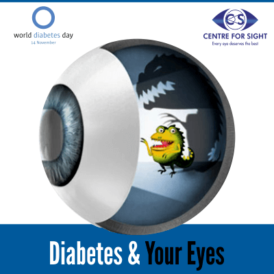
  “World Diabetes Day” was introduced in 1991 by the International Diabetes Federation and the World Health Organization with a goal in mind of creating awareness regarding startling statistics of rising Diabetes observed on global scale. This day is observed on November 14 of each year. It has become an active campaign under the International Diabetes Federation each year to address issues facing the global diabetes community. The campaign draws attention to issues of utmost importance due to diabetes in the world keeping them decisively under the spotlight of public, as Diabetes is now recognized as escalating health threat. Diabetes mellitus (DM), simply referred as diabetes, is a cluster of diseases upsetting the body metabolism resulting in the high blood sugar levels, producing the commonly observed symptoms like frequent urination, increased thirst, and increased hunger. A diabetic individual upholds a condition where the extent of blood glucose too prominent, known as hyperglycemia, which is basically due to efficiency of insulin working in body. The reasons can be body isn’t producing requisite insulin, or has non-working cells which are responding inefficiently to the insulin the pancreas produces. Resultant percussion is glucose building in the blood is too elevated, which eventually gets excreted out of body in urine without providing essential energy and growth requirements to the human metabolism. Now point to ponder about adverse effects of Diabetes on parts of bodies, especially eyes! It has been seen that untreated Diabetes can result in severe complications in future. The term which is used for damage to eyes is “diabetic retinopathy” where retinopathy conveys damage to retina. It is a systemic disease which affects major lot of diabetic patients suffering since 10 years or more. But research indicates that at least 90% of these new cases could be reduced if there was proper monitoring of diabetic eyes and their attentive treatment over these years. So it is more than understood that eye care is important for diabetic patients to ensure vision impassive from any deformities. They are prone to get complications from diabetes and leading reason of blindness in age 20 and above. There should be some precautions which should be used for eye-care – 1) Schedule regular appointments with your eye doctor so that any eye problem can be detected early and treated. 2) Maintain control of your blood glucose levels. 3) Blood pressure should be under control. 4) Get your blood cholesterol levels under control. 4) Adopt healthy habits like eating a healthy diet, avoid smoking and exercise regularly. If you have diabetes, you should be more careful about your eyes than normal person because after diabetes, follows blindness. And even if you don’t have diabetes, some care won’t hurt. So proper eye care is a must for a healthy vision lifelong. *The views expressed here are solely those of the author in his private capacity and do not in any way represent the views of Centre for Sight.

Thu May 15 2014
World Retinoblastoma Awareness Week to be observed

India has the highest number of Retinoblastoma (Rb) affected children in the world, with about 1500 new cases reported each year informed Dr. Santosh G. Honavar, while addressing a press conference here in city on the occasion of World Retinoblastoma Awareness Week.

Retinoblastoma is the most common and life-threatening eye cancer in very young children, new born to 3 years of age, generally resulting in removal of the eye. Each year, more than 5000 new cases of retinoblastoma are diagnosed in the World, of which a third (over 1500) are diagnosed in India. Awareness on Retinoblastoma is low and it can be completely curable with life, eye and vision salvage, if detected early. With improved techniques and technologies, survival of children with Retinoblastoma has improved but low awareness is still a cause of concern. If the child is examined early by a child specialist or an eye doctor and referred to an eye cancer specialist for treatment, then the cancer can be controlled and the child’s life, eye and vision can be saved.

Dr. Santosh G Honavar, Ocular Oncologist has been working  for the cause of retinoblastoma for over 15 years and have together treated over 2000 children with retinoblastoma with 95% life salvage, 90% eye salvage, and 85% vision salvage. Dr. Santosh said, “Our time-tested, evidence-based and experience-driven treatment protocols at Centre For Sight for early and advanced intraocular retinoblastoma, orbital retinoblastoma and adjuvant therapy are personalized to each child to optimize the outcome.”, informed Dr. Santosh.

Eye cancer specialist treats retinoblastoma by chemotherapy, laser and brachytherapy (an advanced form of local radiotherapy), focusing on saving the child’s life, salvaging the eye, and protecting the vision. Treatment ensures 95% of the children are saved from death, over 90% have their eye intact, and over 85% have their vision protected. To save the child’s life and prevent the cancer from spreading to other parts of the body, the eye may have to be removed in advanced cases. It is possible then, to fit a life-like artificial eye on the child to ensure normal appearance.

During the World Retinoblastoma Awareness Week May 11-17, efforts are dedicated to generating public awareness about retinoblastoma, the most common and life-threatening eye cancer in very young children, new born to 3 years of age. To provide special care and attention for Retinoblastoma patients, Centre for Sight (CFS) Super Speciality Eye Hospital, Hyderabad has a dedicated team comprising of an ocular oncologist, pediatric oncologist, ophthalmic oncopathologist, expert chemotherapy nurse, and an ocularist, announced Dr. Santosh G. Honavar, Director – Medical Services and Head – Ocular Oncology & Oculoplasty, CFS Hyderabad. The National Retinoblastoma Foundation at the Centre for Sight, Hyderabad is a fully equipped referral centre for cost-effective and comprehensive management of retinoblastoma. The Centre plans to have special programs to promote the awareness on Retinoblastoma during the World Retinoblastoma Awareness Week to be observed from May11-17, 2014.

Some of the symptoms of Retinoblastoma that can be observed are:

  • A white shiny reflex in the child’s eye is a tell-tale symptom.
  • Crossed eyes or ‘squint’, swelling of the eye, and continuous watering are other symptoms.
  • The vision is lost rapidly and the child might frequently bump into objects and get hurt.

Take the child to an eye specialist if:

  • If there is a white glow in the child’s eye
  • If the child’s eyes are misaligned, also called squint or crossed eyes
  • If the eye is red and swollen because of an unknown reason
  • A simple eye examination can help detect retinoblastoma and save your child’s life.

Take a flash photograph of your child:

  • If there is a white shining spot inside the eye, it could be retinoblastoma
  • Rush the child to an eye specialist or an eye cancer expert

Article: World Retinoblastoma Awareness Week to be observed Author: Nalini Ravichandran   |   Mail Today  |   May 15, 2014 | UPDATED 15:13 IST

Publication: Indian Health News

Read more at: http://www.arrowtimesmedia.com/news-events/world-retinoblastoma-awareness-week-to-be-observed-from-may-11-17-2014.html

*The views expressed here are solely those of the author in his private capacity and do not in any way represent the views of Centre for Sight.

Wed May 14 2014
World Retinoblastoma Awareness Week to be observed from May

India has the highest number of Retinoblastoma (Rb) affected children in the world, with about 1500 new cases reported each year informed Dr. Santosh G. Honavar, while addressing a press conference here in city on the occasion of World Retinoblastoma Awareness Week.

Retinoblastoma is the most common and life-threatening eye cancer in very young children, new born to 3 years of age, generally resulting in removal of the eye. Each year, more than 5000 new cases of retinoblastoma are diagnosed in the World, of which a third (over 1500) are diagnosed in India. Awareness on Retinoblastoma is low and it can be completely curable with life, eye and vision salvage, if detected early. With improved techniques and technologies, survival of children with Retinoblastoma has improved but low awareness is still a cause of concern. If the child is examined early by a child specialist or an eye doctor and referred to an eye cancer specialist for treatment, then the cancer can be controlled and the child’s life, eye and vision can be saved.

Dr. Santosh G Honavar, Ocular Oncologist has been working  for the cause of retinoblastoma for over 15 years and have together treated over 2000 children with retinoblastoma with 95% life salvage, 90% eye salvage, and 85% vision salvage. Dr. Santosh said, “Our time-tested, evidence-based and experience-driven treatment protocols at Centre For Sight for early and advanced intraocular retinoblastoma, orbital retinoblastoma and adjuvant therapy are personalized to each child to optimize the outcome.”, informed Dr. Santosh.

Eye cancer specialist treats retinoblastoma by chemotherapy, laser and brachytherapy (an advanced form of local radiotherapy), focusing on saving the child’s life, salvaging the eye, and protecting the vision. Treatment ensures 95% of the children are saved from death, over 90% have their eye intact, and over 85% have their vision protected. To save the child’s life and prevent the cancer from spreading to other parts of the body, the eye may have to be removed in advanced cases. It is possible then, to fit a life-like artificial eye on the child to ensure normal appearance.

During the World Retinoblastoma Awareness Week May 11-17, efforts are dedicated to generating public awareness about retinoblastoma, the most common and life-threatening eye cancer in very young children, new born to 3 years of age. To provide special care and attention for Retinoblastoma patients, Centre for Sight (CFS) Super Speciality Eye Hospital, Hyderabad has a dedicated team comprising of an ocular oncologist, pediatric oncologist, ophthalmic oncopathologist, expert chemotherapy nurse, and an ocularist, announced Dr. Santosh G. Honavar, Director – Medical Services and Head – Ocular Oncology & Oculoplasty, CFS Hyderabad. The National Retinoblastoma Foundation at the Centre for Sight, Hyderabad is a fully equipped referral centre for cost-effective and comprehensive management of retinoblastoma. The Centre plans to have special programs to promote the awareness on Retinoblastoma during the World Retinoblastoma Awareness Week to be observed from May11-17, 2014.

Some of the symptoms of Retinoblastoma that can be observed are:

  • A white shiny reflex in the child’s eye is a tell-tale symptom.
  • Crossed eyes or ‘squint’, swelling of the eye, and continuous watering are other symptoms.
  • The vision is lost rapidly and the child might frequently bump into objects and get hurt.

Take the child to an eye specialist if:

  • If there is a white glow in the child’s eye
  • If the child’s eyes are misaligned, also called squint or crossed eyes
  • If the eye is red and swollen because of an unknown reason
  • A simple eye examination can help detect retinoblastoma and save your child’s life.

Take a flash photograph of your child:

  • If there is a white shining spot inside the eye, it could be retinoblastoma
  • Rush the child to an eye specialist or an eye cancer expert

Article:World Retinoblastoma Awareness Week to be observed from May 

Author: Nalini Ravichandran   |   Mail Today  |   May 14, 2014 | UPDATED 03:02 IST

Publication: Indian Health News

Read more at: http://newssuperfast.com/2014/05/12/world-retinoblastoma-awareness-week-to-be-observed-from-may-11-17-2014/

*The views expressed here are solely those of the author in his private capacity and do not in any way represent the views of Centre for Sight.

Load More Posts
Blog Archive
Appointment Specialist Locate Us Call Us
"I chose Centre for Sight to get rid of my glasses. Their treatment is permanent, has no side effects and gave me the freedom to live to the fullest."
Select Contact Method
Delhi NCR
Rest of India
Find a Specialist
Locate Us
In Delhi / NCR