|
Key Takeaways:
|
Hypertensive retinopathy is eye damage caused by high blood pressure that harms the tiny blood vessels in the retina. The confusion is that vision may feel normal for a long time, so people often miss the early signs, while BP spikes quietly increase the risk of vision loss. In this guide, you’ll learn what it is, why it happens, the common symptoms and stages, and the treatment and prevention steps that work in India.
What Is Hypertensive Retinopathy?
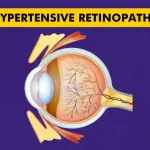
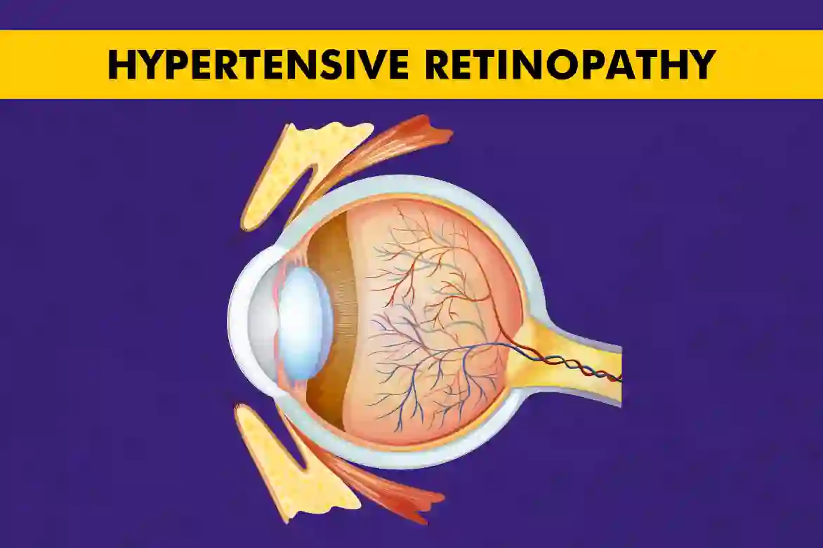
Hypertensive retinopathy is damage to the retina’s tiny blood vessels caused by high blood pressure. Early on, vision can feel normal, but the vessels narrow and leak quietly, and the optic nerve can swell in severe spikes.
An eye exam can detect clues such as narrowed arteries, arteriovenous (AV) nicking, small retinal bleeds, cotton-wool spots, and, in severe cases, papilledema (disc swelling).
Detecting these changes is important because they signal an increased risk of eye problems and a higher risk of heart–brain issues, such as stroke.
Stages of Hypertensive Retinopathy
Hypertensive Retinopathy Grades changes on a 4-step scale (Keith–Wagener–Barker). It runs from mild vessel narrowing to severe damage with optic-nerve swelling.
- Grade 1 (Mild Stage): Subtle, generalized arteriolar narrowing; usually no symptoms.
- Grade 2 (Moderate Stage): Narrowing with AV nicking (arteries compress veins where they cross).
- Grade 3 (Severe Stage): Grade 2 plus retinal hemorrhages, cotton-wool spots, and hard exudates; vision blur.
- Grade 4 (Malignant/Ophthalmic emergency): Grade 3 plus papilledema (swollen optic disc) and marked vessel leakage, needs urgent BP control.
|
Note: You may also hear doctors describe phases, vasoconstrictive, sclerotic, exudative, which reflect how vessels first narrow, then stiffen, then leak if BP stays high. |
Causes and Risk Factors
High blood pressure over time is the main cause of hypertensive retinopathy. Constant high force inside the eye’s tiny arteries makes them narrow, stiff, and leaky, which can lead to bleeding and swelling in the retina.
Good blood pressure control is the core treatment because it lowers the strain on these vessels and reduces the risk of vision problems.
Below are the causes and risk factors for Hypertensive Retinopathy:
- Long-term, uncontrolled hypertension: The longer BP stays high, the greater the vessel injury and the higher the stage of retinopathy.
- Heart–blood vessel disease and high cholesterol: These conditions add stress to retinal circulation and are linked with worse retinal findings and future stroke risk.
- Kidney problems, diabetes, and obesity: Systemic diseases that raise or reflect vascular stress are associated with retinopathy and its progression.
- Smoking and alcohol/tobacco use: Damage blood vessels and increase cardiovascular risk, which travels with retinal risk.
- Age and genetic tendency: Risk rises with age; family and genetic factors influence vessel calibre and BP control.
- Steroid medicines (any form): Can raise BP or interact with eye/vascular health, and should be disclosed to your doctor.
Symptoms of Hypertensive Retinopathy
Early on, most people feel normal. The retina can show changes even when vision seems fine, which is why routine eye exams are important if you have high BP.
Below are the symptoms of Hypertensive Retinopathy:
- Blurred or dim vision: From fluid leakage, small retinal bleeds, or macular swelling; clarity may fluctuate at first.
- Areas of missing or fuzzy detail: Cotton-wool spots and hemorrhages can reduce sensitivity in parts of the retina.
- Headache or eye discomfort: More common when BP is very high or rising quickly.
- Sudden, marked vision drop with severe hypertension: In malignant/grade 4 disease, the optic nerve can swell (papilledema) and the macula can be affected; this is an emergency that needs immediate BP control in the hospital.
Diagnosis of Hypertensive Retinopathy
Doctors confirm hypertensive retinopathy by looking at the back of the eye (fundus) after dilating the pupil.
With a special microscope/light (ophthalmoscopy or slit-lamp), they check for narrowed arteries, arteriovenous (AV) nicking, flame-shaped hemorrhages, cotton-wool spots, hard exudates, and, when blood pressure is very high, swelling of the optic disc (papilledema).
These findings help decide the grade of hypertensive retinopathy and guide follow-up.
If needed, your doctor may add imaging tests to map the damage and track recovery:
- Fluorescein angiography (FA): A dye test that highlights leaks or blocked flow in retinal vessels, especially useful in severe spikes.
- Optical coherence tomography (OCT): A painless scan that shows retinal thickness and macular swelling in detail; OCT angiography (OCTA) can show tiny blood-flow changes without dye.
Because high blood pressure drives the eye changes, checking and documenting systemic BP is part of the diagnosis and ongoing care.
Very high readings with Grade 3–4 eye signs point to a hypertensive emergency and need urgent medical treatment.
Treatment and Management
The main treatment is to lower and control blood pressure safely, which protects the retina and reduces future risk to the heart and brain.
Below are the treatment and management options for Hypertensive Retinopathy:
- Plans usually combine lifestyle changes (DASH-style diet, less salt, regular activity, weight control, no tobacco) and medicines chosen by your physician (e.g., ACE inhibitors, ARBs, calcium-channel blockers, diuretics, beta-blockers).
- If BP is dangerously high with eye findings or other organ symptoms, it is a hypertensive emergency, and treatment must start immediately in the hospital with careful BP lowering.
- Eye-specific care focuses on monitoring and treating complications (for example, macular swelling) while BP is brought under control. With sustained BP control, milder retinopathy improves over months, and your eye team will use exams and imaging to track changes.
- Good follow-up matters: regular blood pressure checks, dilated eye exams, and, when advised, OCT/photography help confirm that vessel changes are settling and vision is stable.
Conclusion
Most cases of hypertensive retinopathy improve when blood pressure is brought under steady control.Eye changes can be silent at first, so regular dilated exams with eye specialists and imaging tests help detect problems early.
In severe spikes, urgent BP treatment protects both sight and overall health. With good BP control, healthy habits, and planned follow-ups, long-term vision can be preserved.
FAQs
Can high BP affect the eyes?
Yes, high BP can damage the retina’s small blood vessels, causing leaks, swelling, and in severe cases, optic-nerve swelling; this is called hypertensive retinopathy.
What Is Hypertensive Retinopathy?
Hypertensive retinopathy is eye damage from long-standing or very high blood pressure that injures the retina’s tiny vessels and can blur or threaten vision.
What are the symptoms of Hypertensive Retinopathy?
Symptoms of Hypertensive Retinopathy may be none at first, then blurred or dim vision, patches of missing detail, and headache; a sudden drop in vision with very high BP needs urgent care.
Can Hypertensive Retinopathy be reversed?
Yes, mild changes improve over months with tight BP control, but severe damage (like scarring or nerve swelling) may leave lasting effects, so early treatment is best.
What is the difference between Hypertensive and Diabetic Retinopathy?
The primary difference between Hypertensive and diabetic retinopathy lies in their causes and appearances: hypertensive retinopathy results from high blood pressure, characterised by vessel narrowing and flame hemorrhages, whereas diabetic retinopathy is caused by high blood sugar, marked by microaneurysms, exudates, and the formation of new, fragile vessels in advanced stages.
How do we differentiate between diabetic and hypertensive retinopathy?
Diabetic retinopathy often involves abnormal blood vessel growth in the retina. It is caused by diabetes, while hypertensive retinopathy is characterised by narrowed or leaking vessels caused by high blood pressure. Both conditions affect the retina but have different underlying causes and patterns of damage.
Can high BP affect the eyes?
Yes, high blood pressure can significantly affect the blood vessels in the eyes, leading to complications like retinal damage. Over time, if untreated, this can result in vision problems or even permanent vision loss.