|
Key Takeaways:
|
Melanoma eye cancer is a rare cancer that starts from pigment-making cells inside or on the surface of the eye, and it can be easy to miss early. The conflict is that many people have no pain and sometimes no symptoms, so diagnosis can happen late or by accident during a routine eye exam.
In this blog, you’ll learn the melanoma meaning in the eye, the causes and symptoms, the types of melanoma, how doctors diagnose it, the main treatment choices, and when you should seek help quickly.
What is Melanoma Of The Eye?
Melanoma is a cancer that starts in melanocytes, the cells that make pigment (colour). When it starts in the eye, it most commonly develops in the uvea (the layer that includes the iris, ciliary body, and choroid), and less commonly on the conjunctiva (the clear “skin” over the white of the eye).
A useful reality check is that eye melanoma is rare. A large SEER trend analysis reported an incidence of 5.6 per million for uveal melanoma, with a 5-year relative survival of 82.8% (1975–2020).
Types Of Melanoma Of The Eye
There are different types of melanoma in and around the eye, and the “type” mostly refers to where it starts. This matters because symptoms, treatment, and spread risk can differ by location.
|
Type (where it starts) |
How common? |
What do people notice? |
Treatment approach |
|
Uveal melanoma(iris/ciliary body/choroid) |
85–90% of ocular melanomas |
Visual symptoms (blur, flashes/floaters) or none early |
Radiation (plaque/proton), surgery in select cases, sometimes observation for very small lesions |
|
Conjunctival melanoma(surface of eye) |
Very rare; incidence about 0.46 per million/year |
Dark spot on the white of the eye that grows/changes |
Surgery + “freeze” treatment; sometimes chemotherapy eye drops |
|
Eyelid skin melanoma (around eye) |
Not “inside-the-eye” melanoma |
Changing mole on eyelid skin |
Managed like skin melanoma (derm/oculoplastics) |
Note: Most uveal melanoma begins in the choroid (about 90%), with smaller proportions in the ciliary body and iris (commonly reported around 6% and 4%, respectively).
Causes Of Melanoma Of The Eye
Doctors don’t find one single cause. Instead, they look at risk factors that make eye melanoma more likely.
- Light eye colour / fair skin / European ancestry: These features show up repeatedly as risk patterns in uveal melanoma research. It does not mean darker eyes are “safe,” but it changes probabilities.
- A pre-existing choroidal nevus (eye mole): Most nevi never become cancer, but the risk of malignant change for a given lesion has been estimated around 1 in 5,000 to 1 in 8,845 in clinical references.
- Ocular or oculodermal melanocytosis (Nevus of Ota): AAO guidance notes the risk of uveal melanoma in people with ocular/oculodermal melanocytosis is estimated around 1 in 400.
- Genetic predisposition (rare, but important): Some families carry higher risk through tumour-predisposition pathway.
Does sunlight cause eye melanoma?
This is the part many people find confusing. UV exposure is a clear driver for skin melanoma, but for uveal melanoma, the link is not as straightforward and is still discussed as a possible factor alongside genetics and pigmentation.
Many clinical findings list UV as a possible contributor, but they don’t present it as the single dominant cause the way it is for skin disease.
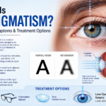
Symptoms Of Melanoma Of The Eye
This condition can be quiet early on, which is why symptoms need to be taken seriously when they appear. Many people notice vision changes or a visible dark spot, but some have no symptoms at first.
- Blurred or distorted vision in one eye: This can feel like “my glasses suddenly aren’t working” on one side.
- Flashes and floaters: These can happen for many harmless reasons too, but new flashes/floaters are still worth checking, especially if one eye feels different.
- A growing dark spot on the iris or conjunctiva: This is one of the most visible clues when the tumour is in the front of the eye.
- Change in pupil shape: A pupil that looks less round can be a warning sign.
- Loss of peripheral vision: Some people notice side vision changes in one eye.
Diagnosis Of Melanoma Of The Eye
Diagnosis starts with an eye specialist looking at the back of the eye through a dilated exam. For uveal melanoma, experienced ocular oncology specialists can diagnose based on examination and ultrasound with very high accuracy, one cancer care review reports 98% accuracy without needing biopsy in many cases.
- Dilated retinal exam + detailed imaging (fundus photography, OCT in many clinics): This shows the lesion’s appearance and whether it is affecting the retina.
- Ocular ultrasound (B-scan): A key test for measuring tumour thickness and internal features.
- Angiography (in select cases): Helps understand blood flow patterns when doctors need more clarity.
- Biopsy and genetic testing (select cases): Not always required to diagnose, but may be used for prognostic testing and risk-stratifying follow-up.
|
Why does the liver get mentioned during follow-up? Uveal melanoma spreads to the liver in most metastatic cases, reported around ~90% of patients with metastasis. Because of that, many follow-up plans focus on liver surveillance. A radiology review describes a common European approach as liver ultrasound every 6 months for 10 years, with CT/MRI if anything looks suspicious. |
Risk Factors for Melanomas of the Eye
With cancer, genetics and certain lifestyle conditions play a role in increasing your risk of developing the disease. This also holds true for melanoma of the eyes. Some of the risk factors for the same are:
-
Genetic mutations passed from parents to children
-
High exposure to UV lights
-
A predisposition to develop many moles
-
Having a light eye colour
-
Being over the age of 45
Melanoma Treatment O The Eye
Treatment depends on location, tumour size, vision potential, and whether there are signs of spread:
- Plaque brachytherapy (eye plaque radiation): A small radiation “plaque” is stitched onto the outside of the eye for a few days to treat the tumour from close range. A plaque series reported 10-year local control of 94%.
- Proton beam therapy: An external radiation technique that can be useful for certain tumour shapes/locations. A proton cohort review reported 10-year tumour control of 87.5% in one large experiment.
- Surgery (including enucleation in selected cases): Removing the eye is reserved for very large tumours, severe pain, or when vision cannot be saved, but decisions are individualized.
- Laser-based treatments (select small tumours): Photocoagulation/thermotherapy concepts in selected situations, for small lesions or adjunct use.
- Conjunctival melanoma treatments: AAO notes treatment can include surgery, freezing therapy, and chemotherapy eye drops for surface lesions.
|
Important: If the cancer has spread? This is harder to treat, but progress is real. For HLA-A*02:01–positive patients with metastatic uveal melanoma, tebentafusp improved survival in a major trial; NCI reports a median overall survival of 21.7 months vs 16 months in the control group. |
Can Melanoma Of The Eye Be Cured?
This is the question most people mean: “Can it be treated early enough to protect life and vision?”
For many patients, the primary eye tumour can be controlled well with radiation or surgery, and the SEER analysis shows a 5-year relative survival of 82.8% for uveal melanoma overall.
The harder part is metastasis risk. A recent review reports distant metastases occur in 25–31% within 5 years, 34–45% within 15 years, and around 50% within 25 years, and the liver is involved in about 90% of metastatic cases.
So, cure depends on stage and biology, but early detection, proper treatment, and structured follow-up make a real difference in outcomes.
When to See a Doctor?
This part matters because many people wait until vision is badly affected.
- New blur or distortion in one eye
- New flashes/floaters that feel unusual or one-sided
- A new or growing dark spot on the iris or on the white of the eye
Treat it as an emergency if you notice:
- Sudden major drop in vision in one eye
- Rapidly worsening side vision loss
- New severe pain with vision change (needs urgent evaluation even if the cause is not melanoma)
Conclusion
Melanoma eye cancer is rare, but it’s one of the reasons routine dilated eye exams can literally save sight. The key idea is simple: the primary tumour is treatable with eye-sparing radiation or surgery, but long-term care also focuses on spread risk, especially to the liver.
If you notice new one-eye blur, flashes/floaters, or a growing dark spot, getting checked early is the safest move.
FAQs
What is melanoma of the eye in simple words?
Melanoma of the eye is a cancer of pigment-making cells that starts inside the eye (in the uvea) or on the eye surface (conjunctiva), and it does not cause symptoms at the start.
What are the main types of melanoma of the eye?
The main types of melanoma of the eye are uveal melanoma (iris, ciliary body, choroid), and conjunctival melanoma, which is much rarer and starts on the eye surface.
What are the early symptoms of melanoma eye cancer?
The early symptoms of melanoma eye cancer are blurred or distorted vision in one eye, flashes/floaters, a growing dark spot on the iris or conjunctiva, a change in pupil shape, or reduced side vision.
How is melanoma eye cancer diagnosed?
Melanoma eye cancer is diagnosed by a dilated eye exam and ocular ultrasound, specialist reviews report experienced ocular oncology clinicians can diagnose uveal melanoma with about 98% accuracy without biopsy in many cases.
Can melanoma of the eye spread, and where does it usually go?
Yes, melanoma of the eye can spread, and when it does, the liver is involved in most metastatic cases (around 90% reported), which is why follow-up plans include regular liver imaging.