|
Key Takeaways:
|
Your retina is delicate, and when it tears, it can set off a chain reaction that may lead to retinal detachment if not treated in time.
The tricky part is that a tear starts with symptoms people brush off, like new floaters or quick flashes, because they don’t feel “serious.”
In this blog, you’ll learn what the retina does, what a retinal tear is, the significant causes, who is most at risk, the warning signs that need urgent help, and the treatments that can prevent bigger damage.
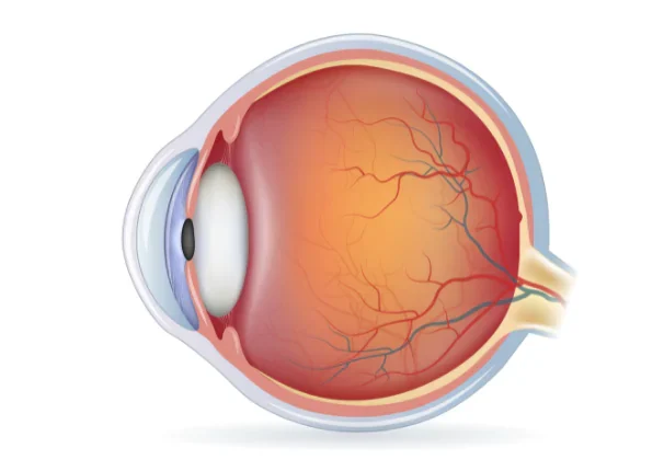
What The Retina Does?
The retina is a thin, light-sensitive layer at the back of your eye that works like a camera sensor.
It converts light into signals that travel through the optic nerve to your brain, helping you see details, colours, and movement.
Because it’s thin and tightly connected to the gel inside the eye (the vitreous), any strong pull on it can create a tear, especially in weak areas.
What Is A Retinal Tear?
A retinal tear is a break in the retina, usually caused by traction, meaning the vitreous gel pulls on the retina hard enough to rip it.
If fluid passes through the tear, it can lift the retina off its base, leading to retinal detachment, which is a vision emergency.
Example: Imagine wallpaper stuck to a wall. If you tug one spot hard, it will rip. Once there’s a rip, moisture can seep behind the wallpaper and start peeling it away, this is similar to how a tear can progress to detachment if untreated.
Causes Of Retinal Tear?
Most retinal tears happen because the vitreous gel changes with age and starts separating from the retina. But there are several casues that increase the pull or weaken the retina.
Posterior vitreous detachment (PVD)
A posterior vitreous detachment happens when the vitreous gel pulls away from the retina as it liquefies and shrinks with age. PVD itself is common, but the pull during separation can create a tear.
How does a tear happen with symptomatic PVD?
- Patient guidance and clinical references commonly state about 10–15% of people with PVD symptoms develop a retinal tear. (The Royal College of Ophthalmologists)
- EyeWiki also notes ~15% of patients with symptomatic PVD have a retinal tear, and the risk jumps much higher (reported 50–70%) if PVD is accompanied by vitreous haemorrhage. (EyeWiki)
- A clinical study of patients with PVD-related symptoms found retinal tears in 14.5%. (National Library of Medicine)
Even if your first exam is normal, some tears can appear later. One large study reported 7.39% of patients with acute symptomatic PVD (without a tear at first visit) developed delayed retinal tears over follow-up. (American Academy of Ophthalmology)
Eye trauma or injury
A hit to the eye (sports injury, accident, blunt trauma) can shake the vitreous and retina and create traction tears.
Trauma-related tears are especially important because they may occur alongside bleeding that blocks a clear view of the retina, so timely assessment matters.
Use of protective eyewear in high-risk sports and workplaces is one of the simplest prevention steps for trauma-related retinal problems.
High myopia (near-sightedness)
With high myopia, the eye is longer, and the retina is more stretched and thinner at the edges. That makes it easier for traction to cause tears.
A review reports the risk of developing retinal detachment is 5–6 times higher in people with high myopia compared with low myopia, one reason doctors watch peripheral retina more closely in high myopes. (National Library of Medicine)
Lattice degeneration (thin/weak peripheral retina)
Lattice degeneration is a peripheral retina condition where the retina becomes thin and weak in patches. It matters because tears can form at lattice borders when the vitreous pulls.
A classic review of prior studies reports lattice degeneration prevalence around 8.0–10.7% in the general population. (ScienceDirect)
Does lattice always cause detachment?
No. Many people with lattice never have problems. But it does raise risk, and long-term data in lattice patients showed cumulative retinal detachment incidence up to 5.3% by age 80 (from holes + tears combined). (National Library of Medicine)
Previous eye surgery
Certain eye surgeries can increase vitreous movement or change traction patterns. Cataract surgery is a common example in population studies.
- A review on post-cataract retinal detachment notes reported incidence ranges from 0.2% to 3.6% depending on follow-up time and patient factors. (National Library of Medicine)
- Registry-based analysis has highlighted that lattice degeneration is a significant risk factor for retinal tear/detachment after cataract surgery. (American Academy of Ophthalmology)
This doesn’t mean cataract surgery is “unsafe”, it’s generally very successful, but it explains why retina risk factors are taken seriously during follow-ups in certain patients.
Inflammation and eye disease
Inflammation inside the eye (uveitis and related conditions) can alter the vitreous and its attachments, increasing traction risk in some cases.
It’s not the most common pathway, but it becomes important when symptoms change quickly or when there’s recurrent inflammation.
Family or personal history
If you’ve had a retinal tear/detachment in one eye, or close relatives have a strong history, your baseline risk can be higher, especially if combined with myopia or lattice.
This is why doctors recommend closer monitoring for the fellow eye in certain high-risk situations.
Myths Vs Facts about Retinal Tear
Myth 1: Reading in dim light causes retinal tears.
Fact: Dim light causes eye fatigue or headaches, but retinal tears are usually due to vitreous traction (often PVD) or retina weakness (myopia/lattice), not reading conditions.
Myth 2: If floaters are common, they’re never urgent.
Fact: Many floaters are harmless, but sudden new floaters, especially with flashes, can signal PVD with a tear. Retina specialists advise prompt evaluation for new symptoms.
Myth 3: A retinal tear always causes pain.
Fact: Retinal tears are painless. People notice visual symptoms (floaters, flashes, shadow/curtain), which is why the symptoms matter so much.
Warning Signs That Suggest A Retinal Tear
Retina societies and major eye-health sources consistently flag these symptoms:
- Sudden onset or burst of floaters (described like pepper or cobwebs).
- Flashes of light (photopsia), especially new and repeated.
- A shadow, curtain, or missing side vision (more suggestive of detachment).
When is it an emergency?
These are the patterns doctors worry about because a tear can turn into a detachment. Seek urgent eye evaluation the same day if you have:
- New floaters plus flashes.
- Any curtain/shadow or sudden loss of side vision.
- Any drop in vision along with these symptoms.
How Are Retinal Tears Diagnosed?
- Dilated retinal examination to look for tears, holes, and early detachment.
- If the retina view is blocked by bleeding, an ocular ultrasound will be used to help detect a tear or detachment.
Treatment for Retinal Tears
If a tear is caught before it becomes a detachment, the outlook is very good.
- Laser photocoagulation (laser retinopexy):
Creates tiny burns around the tear, forming a scar “seal” that helps prevent fluid from getting under the retina.
A recent review summarised that retinopexy of symptomatic horseshoe tears is about 93–96% effective in preventing rhegmatogenous retinal detachment from those tears. (ScienceDirect)
- Cryotherapy (cryopexy):
Freezes around the tear from the outer wall of the eye, also creating a sealing scar; used when the tear is hard to reach or visibility is limited.
Why follow-up still matters: Even after a successful laser, some people develop new tears later, especially if they have high-risk features, so your doctor will schedule repeat checks based on your risk profile and symptoms.
Can Retinal Tears Be Prevented?
You can’t prevent every tear (because aging changes in the vitreous are natural), but you can reduce risk and catch problems early.
- Don’t ignore new flashes/floaters: A timely exam is the biggest “prevention” of detachment. PVD resources advise getting checked when symptoms begin.
- Protect your eyes from injury: Use protective eyewear for sports and hazardous work.
- Know your risk profile: If you have high myopia or lattice degeneration, ask your eye doctor how often you need dilated retina checks.
- After eye surgery: follow your surgeon’s follow-up advice carefully, especially if you are highly myopic or have lattice.
Conclusion
The significant causes of retinal tears come back to two themes: the vitreous pulling on the retina and a retina that’s more vulnerable (high myopia, lattice degeneration, past surgery, trauma, or inflammation).
Because tears are painless, your best protection is recognizing the warning signs early, especially sudden floaters, flashes, or a curtain-like shadow, and getting a dilated retina exam quickly.
When treated promptly with laser or cryotherapy, most retinal tears can be sealed effectively and the chance of progressing to detachment drops dramatically.
FAQs
What are the most significant causes of retinal tears?
The most significant cause of retinal tears is posterior vitreous detachment (PVD), where the vitreous pulls away and can rip the retina; other major causes include trauma, high myopia, lattice degeneration, and higher-risk situations after eye surgery.
Can posterior vitreous detachment (PVD) cause a retinal tear?
Yes, posterior vitreous detachment (PVD) can cause a retinal tear, while many PVDs are uncomplicated, multiple clinical sources report roughly 10–15% of symptomatic PVD cases involve a retinal tear, and the risk is higher when there is vitreous haemorrhage.
What are the warning signs of a retinal tear?
The warning signs of a retinal tear are sudden new floaters (many at once), flashes of light, and any shadow/curtain in vision are the classic signs that should trigger urgent evaluation because they can indicate a tear or detachment.
How are retinal tears treated, and is laser effective?
Most retinal tears are treated with laser retinopexy or cryotherapy, and evidence summaries report retinopexy for symptomatic horseshoe tears is ~93–96% effective at preventing detachment from those tears when done promptly.
Can retinal tears heal on their own without treatment?
No, retinal tears cannot heal on their own without treatment. Leaving it untreated can allow fluid to pass underneath and cause retinal detachment; prompt treatment before detachment generally leads to an excellent prognosis.