Table of Contents
- Understanding High Myopia: More Than Just Nearsightedness
- The Anatomy of the Retina: Why It’s Vulnerable in Myopia
- How High Myopia Contributes to Retinal Tears
- Symptoms of Retinal Detachment Early
- Diagnostic Tools for Early Detection
- Preventive Measures and Treatment Options for High Myopia Patients
- Treatment Options for Tears and Detachment
- Children and High Myopia: Why Pediatric Care Matters
- Conclusion
- FAQs
For people with high myopia, or extreme nearsightedness, blurry distance vision is only the tip of the iceberg. High myopia power isn’t just about thick glasses; it changes the very structure of your eye and increases the risk of serious, sight-threatening complications. Among the most worrying are retinal tears and retinal detachment makes the fact true that yes, high myopia can lead to permanent vision loss if not diagnosed and treated early.
This in-depth explains why patients with high myopia need to be extra vigilant, what symptoms to watch for, and the most effective ways to safeguard their sight.
Understanding High Myopia: More Than Just Nearsightedness
High myopia is defined clinically as a refractive error of -6.00 diopters or worse. While mild myopia simply means you need glasses to see far away, extreme nearsightedness is much more serious because of the way it stretches and thins the eye.
Clinical Facts about High Myopia:
- The eye becomes elongated (longer front to back)
- The retina, the thin, delicate “film” at the back of your eye, is stretched continuously
- The thinning means blood vessels and nerve layers are under constant stress
- It’s a risk factor not just for poor sight, but for sight-threatening diseases
Higher Risks for High Myopes:
- Retinal tears and detachments
- Early eye cataracts
- Macular degeneration, like myopic macular degeneration
This extra risk exists with all types of myopia, whether your myopia is genetic, developed in childhood, or appeared later; the complications are linked to the physical shape of the eyeball, not just your glasses number.
The Anatomy of the Retina: Why It’s Vulnerable in Myopia
The retina is a multi-layered sheet of light-sensitive nerve tissue lining the back wall of your eye. In high myopia:
- The retina gets thinner as it stretches over the elongated eye
- The peripheral (side) retina is especially fragile
- Areas like the vitreous base (where the gel attaches) and the ora serrata are prone to weak spots and holes
- Lattice degeneration, a thinning and abnormality in the peripheral retina, is much more common
- Thin areas can tear or develop holes with very little warning
These changes drastically increase the lifetime risk of both retinal tears and detachment.
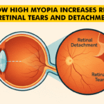
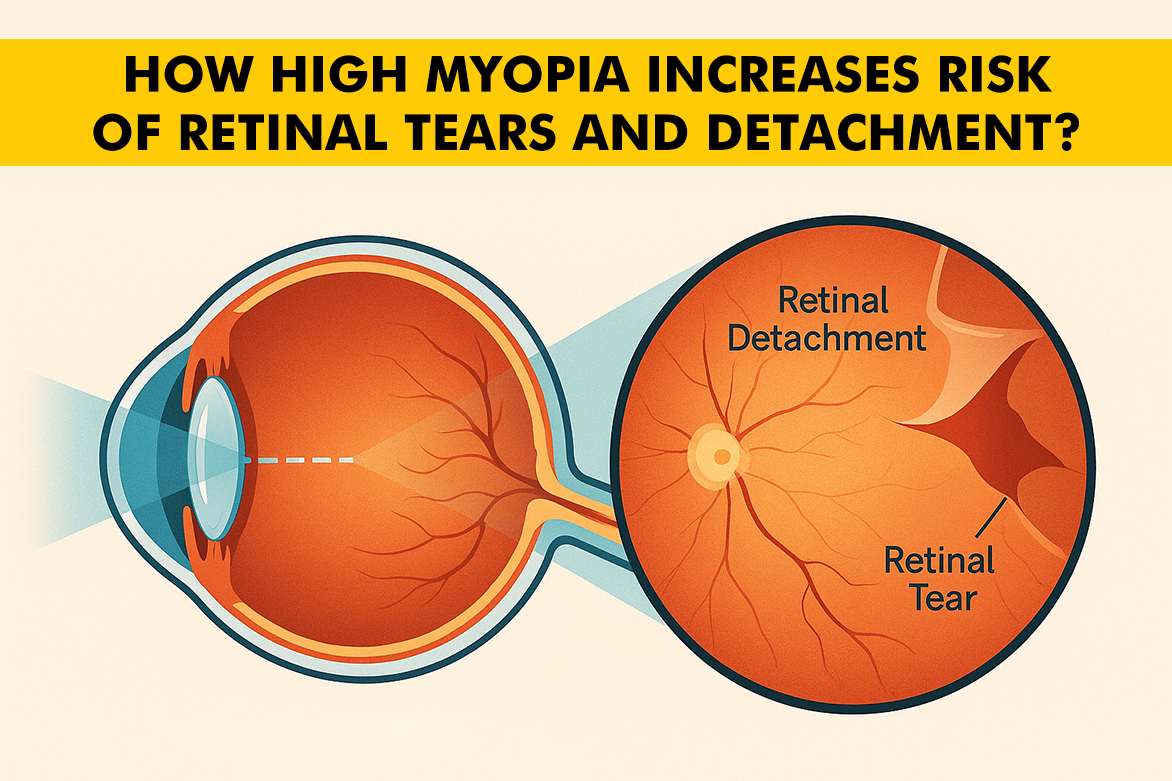
How High Myopia Contributes to Retinal Tears
Among all the types of myopia, high myopia makes the retina more prone to tears because:
- As the vitreous gel in the center of the eye changes with age, it can tug more on a stretched, thin retina
- Sudden movements, trauma, or even regular “vitreous syneresis” (gel liquefaction) put more stress on weak points
- Lattice degeneration (seen in up to 40% of high myopes) frequently precedes retinal tears
- People with high myopia are 10 times more likely to experience retinal detachment than people with normal vision
- Young adults and even children with high myopia can suffer retinal tears, emphasizing the need for regular exams at all ages
Symptoms of Retinal Detachment Early
Prompt recognition is critical for preserving vision. The first symptoms can be very subtle but progress quickly!
- Sudden appearance of many new floaters (tiny spots or wiggly lines drifting in your sight)
- Flashes of light (like lightning or camera flashes), especially at the edge of vision
- A “curtain” or shadow passing across your field of vision
- Rapid decline in vision or a “grey-out” in one area
- Sometimes brief, sharp pain (though detachment itself usually isn’t painful)
If you notice any of these symptoms, especially with high myopia, seek an urgent eye exam; every hour counts!
Diagnostic Tools for Early Detection
Preventing serious retinal problems starts with proactive monitoring with tests like:
- Comprehensive Dilated Eye Exam: Allows the ophthalmologist to see the entire retina, even the far periphery.
- Optical Coherence Tomography (OCT): Gives high-resolution images of retinal layers and can detect subtle holes, thinning, or fluid under the retina.
- Retinal Photography: Documents changes and can be compared over time.
- Ultrasound Imaging: Checks the retina for clouding or bleeding that blocks the view.
- Scleral Indentation: A special manual technique to find tears hiding in folds.
For high myopes, annual or bi-annual exams are essential, more frequent if new symptoms develop.
Preventive Measures and Treatment Options for High Myopia Patients
- Schedule regular retina checks, not just basic vision tests.
- Wear protective eyewear for contact sports or risky jobs.
- Limit eye strain: Follow the “20-20-20” rule when using screens (every 20 minutes, look 20 feet away for 20 seconds).
- Manage conditions like diabetes and blood pressure.
- Atropine drops and special contact lenses are being studied to slow myopic progression in children
- High-dose antioxidants or vitamin supplementation may protect the retina in some cases (evidence is still developing)
Treatment Options for Tears and Detachment
If a Tear Is Detected Early:
- Laser Photocoagulation: Creates a barrier scar around the tear using focused laser beams. Quick and minimally uncomfortable.
- Cryotherapy: Freezes the area to seal a break without entering the eye.
If the Retina Has Detached:
- Vitrectomy: Surgery to remove the pulling vitreous and replace it with balanced saline, air, or gas.
- Scleral Buckling: A flexible band is placed around the eye to relieve pulling and help the retina reattach.
- Pneumatic Retinopexy: A gas bubble is injected to flatten the retina, followed by laser or cryo.
Clinical Outcomes:
Over 90% of retinal detachments can be successfully reattached if treated promptly; vision recovery depends on speed of intervention and whether the macula (central retina) was affected.
Children and High Myopia: Why Pediatric Care Matters
Children with high myopia are at higher risk of early retinal changes and need regular exams, even if symptoms are absent. Special attention is given to rapid shifts in prescription or a sudden loss of vision. Educate young patients and their caregivers about warning signs.
Living with High Myopia: Lifelong Strategies
- Embrace yearly (or more frequent) retina check-ups.
- Stay alert for symptoms, such as flashes & floaters, and teach family/friends what to look out for; many detachments are found because others notice a change.
- Maintain a healthy lifestyle: don’t smoke, keep your blood sugar in check, and stay active.
Conclusion
High myopia brings much more than the inconvenience of strong glasses; it’s a lifelong condition with real risks to your sight. Early detection, regular eye specialist check-ups, and fast treatment of any changes are your best tools for preserving vision. Whether it’s a routine check or a sudden warning sign, never ignore symptoms; your retina is precious, and with modern eye care, you can keep it healthy for many years to come.