Table of Contents
- What Is the Retina?
- How Many Layers Does the Retina Have?
- 10 Layers of Retina
- Layers of Retina Diagram
- Layers of Retina Mnemonic
- Functions of Retina Layers
- Common Disorders Affecting Retina Layers
- When Should You Consult an Eye Specialist?
- FAQs
The human eye is often compared to a camera, and if the lens is the focusing mechanism, the retina is undoubtedly the film or digital sensor. To understand how we perceive the world, from the vibrant colours of a sunset to the fine print in a book, we must look closely at the layers of the retina.
The retina is a thin, light-sensitive tissue lining the back of the eye. While it may appear as a single membrane to the naked eye, microscopic examination reveals a highly organised architectural marvel. Understanding the 10 layers of retina is not just an academic exercise; it is the foundation for diagnosing and treating sight-threatening conditions like macular degeneration and diabetic retinopathy.
What Is the Retina?
The retina is the innermost, light-sensitive layer of the eye. Its primary job is to convert light energy into electrical signals, which are then sent to the brain via the optic nerve to be interpreted as images. It is essentially an extension of the central nervous system.
Structurally, the retina is positioned between the choroid (the vascular layer providing nourishment) and the vitreous humour (the clear gel filling the eyeball). Any disruption in the retina layers can lead to significant visual impairment, making it one of the most clinically studied parts of the human body.
How Many Layers Does the Retina Have?
A common question in ophthalmology is: how many layers does the retina have? The answer is 10 distinct layers. These retina ten layers are arranged from the outermost (closest to the choroid) to the innermost (closest to the vitreous).
Each layer has a specialised role, ranging from light absorption and signal processing to structural support and waste management.
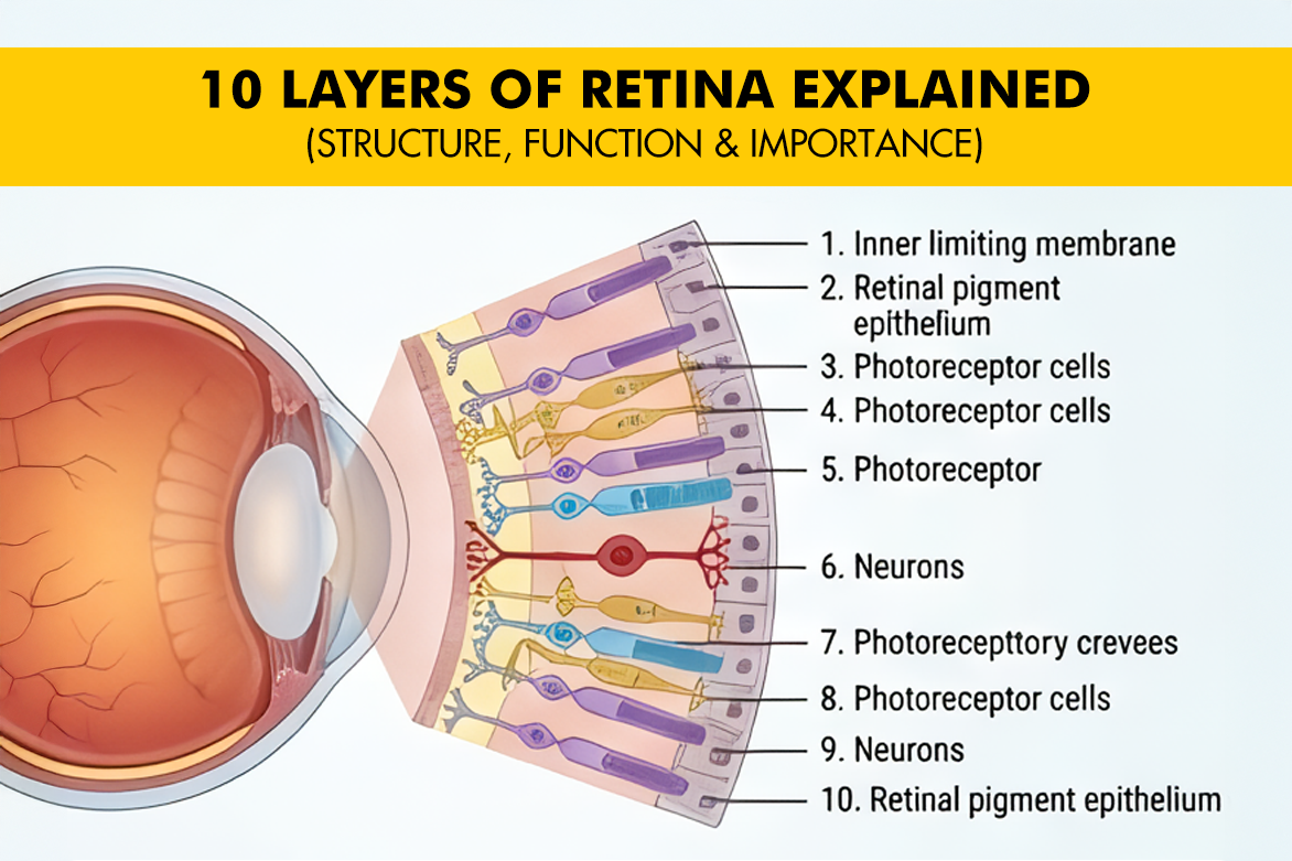
10 Layers of Retina
To understand how vision works, let’s explore the 10 layers of retina in order, starting from the outside (nearest the choroid) and moving inward.
- Retinal Pigment Epithelium (RPE)
The RPE is a single layer of hexagonal cells. It is the outermost layer and is crucial for the health of the photoreceptors.
Function: It absorbs scattered light to prevent glare, transports nutrients from the choroid to the retina, and removes metabolic waste.
Clinical Relevance: Dysfunction in the RPE is a primary factor in Age-Related Macular Degeneration (AMD).
- Layer of Rods and Cones (Photoreceptor Layer)
This is where the “magic” of vision begins. This layer contains the outer and inner segments of photoreceptor cells.
Rods: Responsible for vision in low light (scotopic vision) and peripheral vision.
Cones: Responsible for colour vision and high-definition central vision (photopic vision).
Function: Converting light into chemical energy.
- External Limiting Membrane (ELM)
The ELM isn’t a true “membrane” but rather a row of junctional complexes that link photoreceptors to Müller cells (supportive glial cells).
Function: It provides structural integrity and acts as a mechanical barrier.
- Outer Nuclear Layer (ONL)
This layer contains the cell bodies (nuclei) of the rods and cones.
Function: The thickness of this layer is often measured in clinical scans to determine the density and health of a patient’s photoreceptors.
- Outer Plexiform Layer (OPL)
This is a “synaptic” layer. Here, the extensions of the rods and cones connect with the dendrites of bipolar and horizontal cells.
Function: Initial processing of visual signals occurs here as information passes from photoreceptors to the next level of neurons.
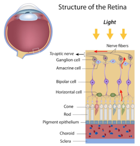
- Inner Nuclear Layer (INL)
The INL is the “processing center.” It contains the cell bodies of several different types of neurons:
Bipolar Cells: Transmit signals from photoreceptors to ganglion cells.
Horizontal Cells: Help the eye adjust to see well under both bright and dim light conditions.
Amacrine Cells: Involved in complex signal processing, such as detecting movement.
- Inner Plexiform Layer (IPL)
Similar to the OPL, this is a synaptic zone. It is where the bipolar cells connect with the ganglion cells. It is here that “ON” and “OFF” pathways are refined, helping the brain distinguish between light and dark boundaries.
- Ganglion Cell Layer (GCL)
This layer contains the cell bodies of the ganglion cells, the final output neurons of the retina.
Clinical Relevance: This layer is particularly sensitive to increased intraocular pressure. Damage here is a hallmark of Glaucoma.
- Nerve Fiber Layer (NFL)
The axons of the ganglion cells travel across the retina in this layer to gather at the optic disc.
Function: These fibres bundle together to form the Optic Nerve.
Clinical Importance: Changes in NFL thickness are a key indicator for early glaucoma detection using OCT technology.
- Internal Limiting Membrane (ILM)
The ILM is the innermost boundary of the retina, formed by the basement membrane of Müller cells.
Function: It separates the retina from the vitreous humour. Surgeons often “peel” this layer during certain vitreoretinal surgeries to treat macular holes.
Layers of Retina Diagram
A diagram of a retina with all its layers is essential for visualising how these microscopic structures stack together. Clinically, doctors use Optical Coherence Tomography (OCT) to produce a high-resolution, cross-sectional “diagram” of a patient’s own retina. By examining these layers, an ophthalmologist can determine which layer is affected. For example, fluid under the RPE may indicate “wet” AMD, while thinning of the Nerve Fibre Layer suggests progression of glaucoma.
Layers of Retina Mnemonic
Memorizing the retina layers can be challenging for students and healthcare professionals alike. A popular layers of retina mnemonic helps remember the order from the Outermost to Innermost:
“Really Reds Eat Out Often; Instead, I Generally Need Icecream.”
- R – Retinal Pigment Epithelium
- R – Rods and Cones
- E – External Limiting Membrane
- O – Outer Nuclear Layer
- O – Outer Plexiform Layer
- I – Inner Nuclear Layer
- I – Inner Plexiform Layer
- G – Ganglion Cell Layer
- N – Nerve Fibre Layer
- I – Internal Limiting Membrane
Functions of Retina Layers
| Layer | Name | Primary Function |
| 1 | RPE | Nutrient transport & waste removal |
| 2 | Photoreceptors | Light detection (Rods & Cones) |
| 3 | ELM | Structural support |
| 4 | ONL | Nuclei of Rods/Cones |
| 5 | OPL | Synapse (Photoreceptor to Bipolar) |
| 6 | INL | Signal processing (Bipolar/Amacrine/Horizontal) |
| 7 | IPL | Synapse (Bipolar to Ganglion) |
| 8 | GCL | Output neuron cell bodies |
| 9 | NFL | Axons forming the Optic Nerve |
| 10 | ILM | Boundary with Vitreous |
Common Disorders Affecting Retina Layers
When one or more of the layers of retina are compromised, symptoms can vary significantly:
- Metamorphopsia (Distorted Vision): Often caused by fluid or deposits (drusen) disrupting the smooth alignment of the RPE and Photoreceptors.
- Retinal Detachment: Occurs when the neurosensory retina separates from the underlying tissue, leading to sudden flashes and floaters.
- Diabetic Retinopathy: Results from diabetes-related damage to retinal blood vessels, causing leakage, bleeding, and progressive vision loss if left untreated.
- Visual Field Loss: Damage to the Nerve Fibre Layer or Ganglion Cell Layer can cause “blind spots” in the peripheral vision.
- Floaters and Flashes: Often occur when the Vitreous pulls on the Internal Limiting Membrane, potentially leading to a retinal tear.
When Should You Consult an Eye Specialist?
Because the layers of retina are so thin and fragile, early detection of damage is critical. You should schedule a comprehensive eye exam if you experience:
- Sudden blurred or distorted vision.
- The appearance of many new floaters or flashes of light.
- A “curtain” or “shadow” falling over your field of vision.
- Difficulty seeing at night.
At Centre For Sight, we utilise world-class diagnostic technology to examine every layer of your retina. Early diagnosis is the best way to protect your gift of sight.