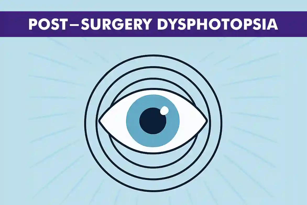
Cataract surgery is a common procedure aimed at restoring clear vision. However, some patients experience unwanted visual disturbances post-surgery, known as dysphotopsia. These effects can range from mildly annoying to significantly disruptive, affecting daily activities and overall quality of life. Understanding these disturbances is crucial for patients to manage expectations and seek appropriate care. This blog will explore the phenomenon of post-surgery dysphotopsia, its types, symptoms, causes, and the available management and treatment options, providing a comprehensive guide for those affected.
Understanding Dysphotopsia: What Is It?
Dysphotopsia refers to the visual disturbances that some people experience after undergoing cataract surgery. These disturbances are often linked to the intraocular lens (IOL) that is implanted during the procedure to replace the eye’s natural lens. Dysphotopsia is generally divided into two main types: positive dysphotopsia and negative dysphotopsia. Positive dysphotopsia involves seeing extra light effects, such as glare or halos, which can be distracting or uncomfortable. On the other hand, negative dysphotopsia is characterized by the perception of dark areas or shadows, often appearing in the peripheral vision, which can be unsettling and may interfere with normal vision.
Common Types of Dysphotopsia: Positive and Negative
Positive dysphotopsia often presents as visual disturbances such as glare, halos, or starbursts around lights. These effects are particularly noticeable in low-light conditions or at night, making activities like night driving or walking in dimly lit areas more challenging. The intensity of these light effects can vary, sometimes causing discomfort or distraction. Additionally, the type of intraocular lens (IOL) used during surgery can influence the severity of positive dysphotopsia, with certain designs being more prone to causing these symptoms.
Negative dysphotopsia, on the other hand, is characterized by the perception of dark shadows or crescents in the peripheral vision. These shadows can appear as dark arcs or bands, often leading to a sensation of something obstructing the side vision. This type of dysphotopsia can be more difficult to diagnose and treat due to its subtle and subjective nature. Factors such as the positioning of the IOL and the patient’s unique eye anatomy can contribute to the occurrence of negative dysphotopsia. It may also require more specialized interventions to address effectively.
Common Symptoms of Post-Surgery Dysphotopsia
- Glare: Bright light reflections that can cause discomfort and make it hard to see clearly.
- Halos: Rings of light that appear around light sources, often noticeable at night.
- Starbursts: Radiating lines or spikes of light around bright objects, affecting visual clarity.
- Dark Shadows: Perceived dark areas in the field of vision, often in the peripheral view.
- Crescents: Curved shadows or arcs that can appear at the edges of vision.
These symptoms can lead to difficulties with activities such as night driving, reading, or tasks requiring detailed vision.
Causes of Dysphotopsia After Cataract Surgery
- Dysphotopsia often results from the interaction of light with the edges of the intraocular lens (IOL) implanted during cataract surgery.
- The design of the IOL can contribute to dysphotopsia if it has sharp edges that refract light, creating visual disturbances.
- The material of the IOL may also cause dysphotopsia, as certain materials can reflect or scatter light differently, leading to unwanted visual effects.
- The positioning of the IOL is crucial; improper alignment can cause light to enter the eye at unusual angles, resulting in dysphotopsia.
- The patient’s unique eye anatomy can influence dysphotopsia, as variations in eye structure may affect light interaction with the IOL.
- The surgical technique used during the procedure can impact the outcome; incorrect placement of the IOL can increase the risk of dysphotopsia.
Management and Treatment Options for Dysphotopsia
Initial management strategies for dysphotopsia focus on a conservative approach, beginning with careful observation and providing reassurance to patients. This is because, in many cases, the brain can gradually adapt to the new visual inputs, leading to a natural resolution of symptoms over time. Patients are encouraged to monitor their symptoms and maintain regular follow-ups with their ophthalmologist to track any changes or improvements.
For those experiencing persistent symptoms that do not improve with time, more proactive treatments may be necessary. One common non-invasive option is the use of anti-glare glasses, which can help reduce the intensity of light disturbances and improve visual comfort, especially in bright environments. Additionally, if the dysphotopsia is linked to the positioning of the intraocular lens (IOL), a surgical adjustment may be considered. This involves repositioning the IOL to optimize its alignment within the eye, potentially alleviating the unwanted visual effects.
In more severe cases, where other interventions have not provided relief, replacing the IOL with a different type may be recommended. This option is typically considered when the design or material of the original IOL contributes significantly to the dysphotopsia. The replacement IOL is carefully selected to minimize the risk of similar issues, taking into account the patient’s specific eye anatomy and visual needs. It is crucial for patients to discuss all available options with their healthcare provider to determine the most suitable course of action based on their individual circumstances.
When to Seek Professional Help for Dysphotopsia Symptoms
Patients should seek professional help if dysphotopsia symptoms persist beyond a few weeks post-surgery or if they significantly impact daily activities.
Consulting an ophthalmologist is crucial for a thorough evaluation and to discuss potential treatment options tailored to the specific type and severity of dysphotopsia.
Conclusion
While post-surgery dysphotopsia can be a challenging and frustrating experience, understanding its causes and available treatments can help manage and alleviate symptoms.
If you are experiencing unwanted visual effects after cataract surgery, don’t hesitate to seek professional advice to explore the best options for your situation.
FAQs
The duration of dysphotopsia varies; some patients may see an improvement in a few weeks, while others may experience symptoms for longer. In many cases, dysphotopsia can resolve on its own as the brain adapts to the new visual inputs.
Certain types of IOLs, particularly those with sharp edges or multifocal designs, may be more likely to cause dysphotopsia. It is essential to discuss these risks with your surgeon before the procedure.
For persistent dysphotopsia, treatments range from non-invasive options like anti-glare glasses to surgical interventions such as IOL repositioning or replacement.
Consult a doctor if visual effects significantly impact your quality of life or if you have concerns about your post-surgery recovery. Early intervention can help address and manage these symptoms effectively.


