Key Takeaways
|
Eye tumour is a broad term for an abnormal growth in or around the eye, and not every tumour is cancer.
Many people panic when they hear the word “tumour,” but some eye tumours are harmless, while others need urgent care to protect vision and health.
In this blog, you will learn what an eye tumour is, the common types, warning signs, diagnosis tests, and how eye tumour treatment is planned.
What is an Eye Tumor?
An eye tumour is an abnormal mass of cells that grows in the eye itself or in nearby structures such as the eyelid, conjunctiva, orbit, retina, or uvea.
It can be benign (non-cancerous) or malignant (cancerous), and it can start in the eye or spread there from another part of the body.
In adults, the most common cancer that starts in the eye is ocular melanoma, while in children, retinoblastoma is the most common primary intraocular malignancy.
For a normal reader, the easiest way to understand this is simple: a tumour is like an abnormal lump or growth. But where it sits matters a lot. A small benign spot on the conjunctiva is very different from a tumour growing in the retina, the choroid, or the orbit behind the eye.
Types of Eye Tumors
Because “eye tumour” covers many conditions, it helps to group them by where they start and whether they are benign or malignant.
| Type | Where does it start? | Benign or malignant | More common in? |
| Conjunctival nevus | Clear surface tissue over the white part of the eye | Benign | Children and young adults |
| Choroidal nevus | Pigmented layer inside the eye | Benign but watched for change | Adults |
| Uveal/ocular melanoma | Uvea, often choroid | Malignant | Adults |
| Retinoblastoma | Retina | Malignant | Young children |
| Conjunctival melanoma | Eye surface | Malignant | Adults |
| Ocular surface squamous neoplasia | Conjunctiva/cornea surface | Pre-cancerous to malignant | Adults |
| Lymphoma/metastasis | Inside eye or orbit | Malignant | Adults |
Causes of Eye Tumor
There is no single cause behind every eye tumour. The reason depends on the tumour type, the person’s age, and whether the growth began in the eye or spread there from somewhere else.
- Genetic changes: Retinoblastoma happens because of changes in the RB1 gene. In hereditary cases, the gene change is present in all body cells, which is why family history matters.
- Pigment-cell cancer changes: Ocular melanoma starts in melanocytes, the pigment-making cells of the eye in the uvea.
- Skin and eye colour-related risk: Older age, fair skin, and light colored eyes increase the risk of intraocular melanoma.
- Spread from another cancer: Some eye tumours are not primary eye cancers at all. They are metastases, meaning cancer from another part of the body has spread to the eye. Choroidal metastases are the most common intraocular tumours overall.
- Surface damage and abnormal cell growth: Ocular surface tumours can be linked with ultraviolet exposure, abnormal surface-cell changes, and in some cases HPV or immune problems, although not every patient has a clear trigger.
Eye Tumor Symptoms
Symptoms vary a lot because some tumors sit quietly for a long time, while others disturb vision early.
Intraocular melanoma has no early signs or symptoms and is sometimes found during a routine eye exam. A child with retinoblastoma will not say, “I have blur.” A parent can first notice a white pupil in flash photos, a wandering eye, or one eye that does not seem to track normally.
That is why examples matter here: adults report visual symptoms, but children are diagnosed because someone notices a change in appearance.
| Symptom | What does a person notice? | Tumours where it appear |
| Blurred vision | Vision looks hazy or distorted | Ocular melanoma, retinal tumours |
| Dark spot on iris or conjunctiva | A visible pigmented patch or growth | Iris melanoma, conjunctival melanoma |
| Flashing lights or floaters | Brief light flashes or moving spots | Posterior tumours affecting retina/vitreous |
| Loss of side or central vision | Missing area in vision | Intraocular tumours |
| White reflex in pupil | White glow in photos or light | Retinoblastoma |
| Eye redness or swelling | Eye looks inflamed or puffy | Retinoblastoma, surface tumours, orbital tumours |
| Eye turning outward or inward | Misaligned eyes | Retinoblastoma in children |
| Pain or pressure | Deep discomfort or aching | Larger tumours, secondary glaucoma, orbital disease |
How is Eye Tumour Diagnosed?
Doctors start with a full eye exam, but the exact tests depend on where the tumour is suspected and how deep it lies. A good diagnosis combines eye examination, imaging, and, in selected cases, tissue testing.
-
Slit-lamp biomicroscope
This is a microscope with a bright light used to examine the front of the eye in detail. It helps doctors study surface growths on the conjunctiva, cornea, iris, and front chamber. It is one of the first machines used when a visible or front-of-eye eye tumour is suspected.
-
Indirect ophthalmoscope and dilated fundus exam
This is used after the pupil is dilated so the doctor can look into the back of the eye. It is important for tumours in the retina, choroid, and optic nerve area, especially when the doctor is checking for ocular melanoma or retinoblastoma.
-
Fundus photography
This machine takes a colour photograph of the back of the eye. It helps document the size, colour, and margins of a lesion so the doctor can compare it over time. This is especially useful for follow-up in pigmented lesions and retinal tumours.
-
Optical Coherence Tomography (OCT)
OCT, or optical coherence tomography, uses light waves to create cross-sectional images.
In tumour work-up, it helps show how the retina and nearby tissue are being affected, such as fluid under the retina, retinal swelling, or structural distortion from a mass.
-
Ocular ultrasound
Ultrasound uses sound waves to show the inside of the eye. Ocular ultrasound is especially useful in ocular melanoma because it helps measure the size of the tumour. It is also helpful when the doctor cannot see clearly through the eye because of cataract, bleeding, or other opacity.
-
Fluorescein angiography
This test involves dye and special photographs of the eye’s blood vessels. It helps the doctor study how the lesion is supplied by blood vessels and whether there is leakage or other retinal circulation change around the tumour.
-
MRI or CT scan
These are not routine for every eye growth, but they become important when the doctor needs to check whether the tumour involves the orbit, surrounding bone, optic nerve, or brain. CT uses X-rays to create slices, while MRI is preferred for soft tissue detail.
-
Biopsy
Biopsy means taking a tissue sample. A biopsy is rarely needed to diagnose intraocular melanoma, because many cases can be diagnosed from examination and imaging alone.
But biopsy can still be used selectively when the diagnosis is uncertain, when molecular testing is needed, or when a surface lesion needs pathology confirmation.
Eye Tumour Management
Management means what doctors do after diagnosis, even before active treatment starts. It is not always the same as treatment.
Some tumours are watched, some are treated quickly, and some need combined care with oncology, retina, orbit, or paediatric teams.
| Management step | Why is it done? | Note |
| Observation with regular imaging | Not every tumour needs immediate treatment | Small stable benign lesions, some small melanomas under observation |
| Vision monitoring | Tracks whether the lesion is affecting sight | Macular or optic-nerve area lesions |
| Systemic work-up | Checks whether the tumour is linked to a body-wide cancer or syndrome | Metastasis, hereditary retinoblastoma |
| Multidisciplinary referral | Brings in retina, ocular oncology, paediatric oncology, or radiation teams | Malignant or complex cases |
| Pain and pressure control | Manages symptoms while planning definitive care | Secondary glaucoma, large tumours |
| Counselling and genetic advice | Helps families understand risk and follow-up | Retinoblastoma and hereditary syndromes |
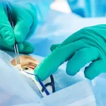
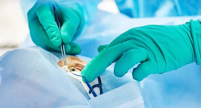
Eye Tumour Treatment
The right eye tumour treatment depends on the exact tumour, its size, its location, whether it is benign or malignant, whether it has spread, and how much vision can still be saved.
That is why treatment for one patient can be “watch and scan,” while another patient needs surgery, radiation, or chemotherapy.
A very important practical point is this: saving life comes before saving the eye, and saving the eye comes before saving every line of vision. Doctors make treatment decisions in that order, especially in malignant tumours in children.
| Treatment | What does it do? | Common tumour situations |
| Observation | Watches a lesion closely without immediate treatment | Small benign lesions, selected small melanomas |
| Laser / photocoagulation / thermotherapy | Uses focused heat or laser energy on the tumour | Selected ocular melanoma or retinoblastoma cases |
| Radiation therapy | Uses targeted radiation to destroy tumour cells | Ocular melanoma, some retinoblastoma cases |
| Surgery to remove the tumour | Removes a localised growth while trying to preserve the eye | Some surface or orbital tumours |
| Enucleation | Removes the whole eye when needed for life or safety | Advanced retinoblastoma, large painful tumours |
| Chemotherapy | Kills or shrinks cancer cells | Retinoblastoma, lymphoma, metastatic disease |
| Targeted therapy / immunotherapy | Uses cancer-specific medicines | Selected advanced ocular melanoma cases |
Conclusion
An eye tumors is not one single disease. It can be a harmless growth, a vision-threatening lesion, or a true cancer that needs urgent specialist care.
The most important thing is not to guess from symptoms alone, because the right diagnosis depends on proper examination, imaging, and sometimes pathology. The good news is that modern eye tumour treatment now includes much more than just eye removal, with options such as observation, laser, radiation, chemotherapy, and targeted therapy depending on the case.
If you notice a new eye lump, white reflex, dark spot, unexplained blur, or persistent redness, the safest next step is an exam with an eye specialist without delay.
FAQs
Is an eye tumour always cancer?
No, an eye tumour is not always cancer. Some eye tumours are benign, such as certain nevi or harmless growths, while others are malignant and need cancer treatment. That is why proper diagnosis matters so much.
What are the first symptoms of an eye tumour?
The first symptoms of an eye tumour depends on the type. Some types cause no early symptoms, while others cause blurred vision, a dark spot on the iris, a white pupil in children, eye redness, swelling, floaters, or side-vision change.
Can eye tumour treatment save vision?
Yes, some eye tumour treatments save vision but not always and fully. In many cases, doctors can now save the eye and sometimes useful vision, especially when diagnosis is not delayed. But the chance of saving vision depends on the tumour type, size, location, and how early treatment begins.
Which doctor treats an eye tumour?
A doctor who treats an eye tumour is called an ocular oncologist, retina specialist, orbit specialist, or paediatric eye cancer specialist, depending on the tumour. Many patients are first seen by a general ophthalmologist and then referred to a specialist team.
Can children get an eye tumour?
Yes, children can get an eye tumour. The best-known example is retinoblastoma, which is the most common primary intraocular malignancy in children and affects very young children.