Imagine waking up with diminution of vision, a ‘bloody eye’ and feeling pain as well. Not a pleasant visual to imagine, right? Well, don’t panic right away, pooling of blood in the front part of the eye is a common incidence and is called hyphema. Heave a sigh of relief as it is possible to identify and treat this eye condition.
Symptoms of hyphema that one should not overlook[1]
- Blood inside the eye: A severe or large hyphema can make the entire eye look discolored. The eye fluid aqueous humor in the front part of the eye is replaced by blood. Visual inspection is required to identify a moderate to big-sized hyphema. However, it may not be enough to identify minor pooling of blood in anterior chamber of eye causing pain and other vision-related problems.
- Distorted vision: Distortion or changes in the vision vary according to the severity of hyphema. With obstruction in the pupil, you may only be able to find or detect a small very less amount of light.
- Hypersensitivity to light: Hyphema creates increased sensitivity to light, also known as photophobia. It causes eyes to hurt and may even result in headaches on exposure to bright light.
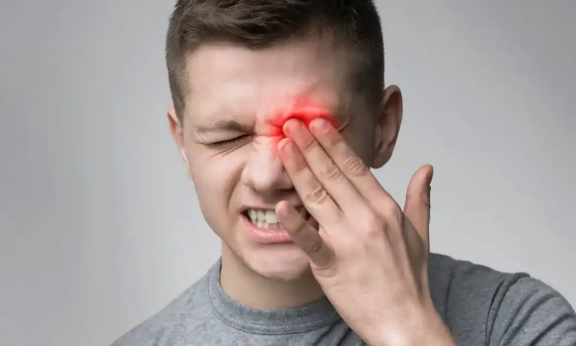
- Eye pain: Accumulation of blood in the front part of the eye between the cornea and iris (anterior chamber) lead to irritation and increased eye pressure which causes pain.
These symptoms of hyphema may keep on worsening based on the degree or grade of hyphema. The condition can progress from grade 0 to grade 4 hyphema. Let us get to know more about the different stages or grades of hyphema:
Grade 0: The pooling of blood can only be identified through a microscope but it is not visible with the naked eye
Grade 1: Around less than one-third of the anterior chamber of the eye is filled with the blood
Grade 2: About half of the chamber is filled with blood
Grade 3: The pooled blood fills more than half of the front part or anterior chamber of the eye, but not completely.
Grade 4: The anterior chamber is filled with blood. If the blood is bright red, the condition is called total hyphema. Whereas, if the hyphema appears dark red, the condition is called 8-ball hyphema.
The sooner you visit an eye specialist for a checkup, the easier it is to get rid of hyphema. This eye condition can arise due to different reasons.
Traumatic hyphema is common and it may occur due to a fall, sports injury, or an accident at the workplace or home. Other causes of hyphema in eye include:
- Unusual blood vessels on the coloured part of the eye called the iris
- Eye infection resulting from catching the herpes virus
- Conditions related to issues in blood clotting such as sickle cell anemia, hemophilia
- Eye cancer
- Problems induced due to intraocular lens or artificial lens implant
Whatever the cause you identify your hyphema with, it is better to seek out medical consultation with an experienced eye doctor. There is no safe alternative to medical diagnosis and treatment. Not taking timely action may cause major eye problems in the long run.
Get a proper diagnosis and steer clear of hyphema complications [2]
It is necessary to discuss your medical history with the doctor to enable them to determine if there is any recent incident or trauma causing bleeding in the eye. The eye doctor will conduct a careful examination to check for deeper signs of the actual problem and advise you the right steps of hyphema management.
- Applanation Tonometry: This test helps in assessing eye pressure. There are different approaches to this eye test, however, the one which makes the corneal area flatten is highly precise. Numbing eye drops are used before the procedure to reduce discomfort. Tonometry involves the use of slit lamp and dye. The doctor uses a machine to get an accurate pressure reading.
- Comprehensive eye checkup: This eye examination is carried out to check a person’s ability to see.
- Slit lamp test: This test enables the doctor to get a clear view of the inside of the eye. A combination of bright light and magnification is used to accurately evaluate the internal structures of the eye.
- CT scan: If hyphema is caused due to trauma, the eye specialist may recommend a CT scan to check if the eye socket is fine.
- Hyphema treatment options [1]
- All hyphema treatment options work on clearing the blood, lowering eye pressure and keeping away recurring bleeding. Most of the cases of hyphema are mild and thus don’t require invasive treatment. A hyphema indicates an underlying injury within the eyeball. Hence, seeking medical advice is necessary. The eye doctor recommends keeping the head of the bed raised to facilitate eye drain. The patient is also asked to wear a protective eyeshield, avoid physical activities, and get eye pressure checked regularly.
- Depending upon the severity of hyphema, the doctor may also prescribe steroid eye drop to subside swelling and inflammation in the eye. Some dilating eye drops may also aid in bringing down the discomfort. While being treated for hyphema, avoid blood thinning medicines like aspirin as it can cause more hemorrhage.
- In severe cases of grade or grade 4 hyphema, eye doctors prescribe beta-blockers, prostaglandins, etc. to cure hyphema with glaucoma. Sometimes, oral medication may also become necessary in serious conditions. When every other alternative fails to give permanent relief, surgical interventions like laser therapy, filtering, drainage tube, or MIGS (minimally invasive glaucoma surgery) may be used.
- Hyphema complications arise due to prolonged pooling of blood in the anterior part of the eye. Without timely medical intervention, you may develop any of the following problems:
- Blood Staining on the cornea: Blood retention in the anterior chamber can result in corneal staining which may further make the vision cloudy.
- Glaucoma: As blood starts pooling inside the eye, it increases the eye pressure or intraocular pressure, which is how glaucoma starts. If left unattended or untreated for a long time, glaucoma may lead to permanent loss of vision.
- Vision loss: Optic damage due to a serious traumatic injury which is non-glaucomatous resulting from long-standing intraocular pressure can cause vision loss when one doesn’t pay immediate attention to hyphema.
- Secondary hemorrhage: A serious traumatic injury may sometimes lead to recurrent bleeding, making the ocular tissue scarred.
Hyphema is one of the common eye problems that can occur to anyone due to an injury or trauma. Getting medical treatment in time prevents serious repercussions and saves vision. Further, being aware of general eye care steps and going for comprehensive eye checkups every six months. To book your consultation and complete eye checkup, visit https://www.centreforsight.net/